Falsification of Davson-Danielli's Model

http://sebiology.weebly.com/uploads/9/0/0/5/90055547/membrane-models-med-1_orig.jpeg
|
Davson and Danielli first proposed their cell membrane model in 1935 based on some specific data concerning phospholipids in cells. However, little information was known about the proteins within the membrane.
The Davson-Danielli Model identified two layers of phospholipids, with tails facing inward, and a layer of proteins on either side of the heads of the phospholipids. (See diagram) |
Falsification Evidence:
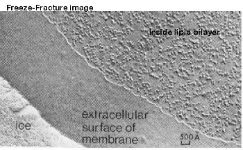
In the 1950s and 1960s, lots of work was done with the cell membrane much of it made possible by the development of X-ray diffraction studies and electron microscopy. Three major pieces of evidence were found to falsify Davson-Danielli's Model.
Freeze-etched eletron micrographys 
https://upload.wikimedia.org/wikipedia/commons/thumb/2/
24/FreezeFracture_final.jpg/550px-FreezeFracture_final.jpg
http://163.178.103.176/tema1g/grupos1/germant1/gatp3/Membrana3_files/ffimage.jpg
|
Structure of membrane proteins Improvements in biochemical techniques allowed proteins to be extracted from the lipid bilayer

https://cdn4.vectorstock.com/i/1000x1000/32/03/membrane-proteins-vector-23523203.jpg
|
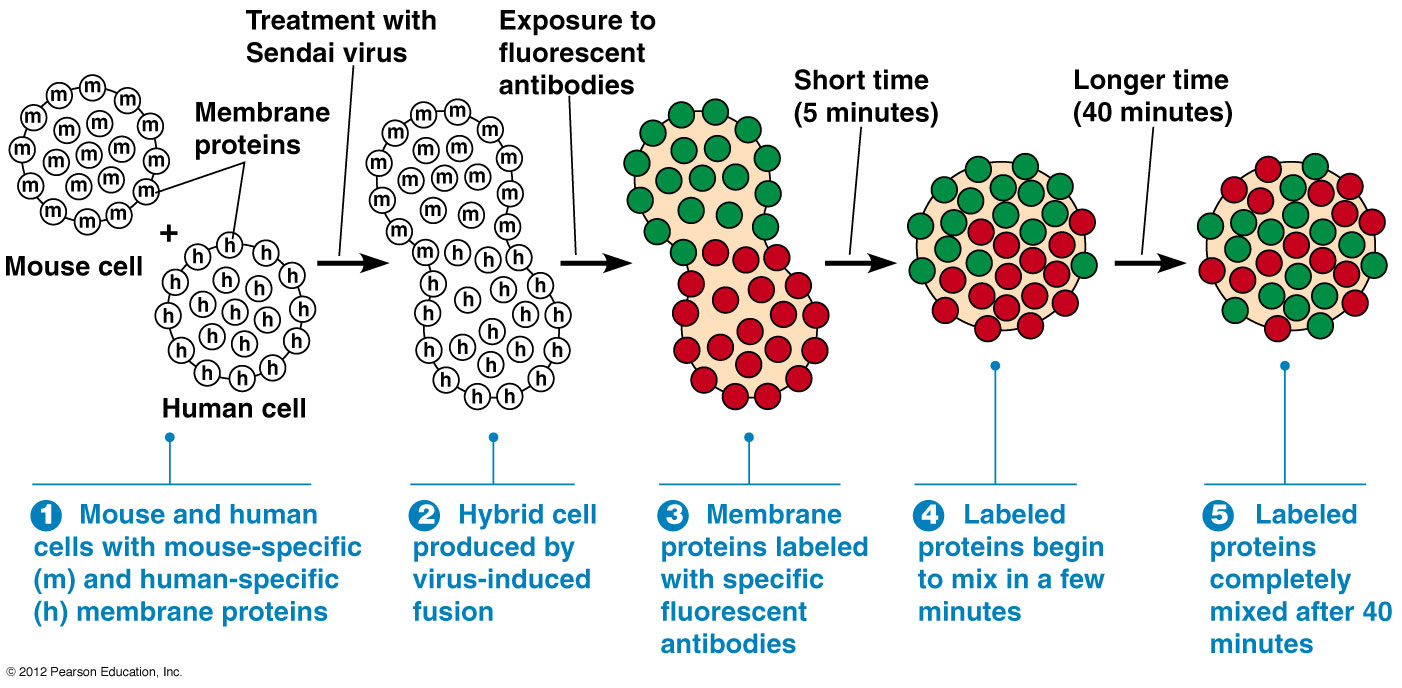
Fluorescent antibody taggingExperiment conducted by Singer- Nicolson in 1972.
http://www.mun.ca/biology/desmid/brian/BIOL2060/BIOL2060-07-08/07_28.jpg
|
Singer- Nicolson's Fluid Mosaic Model
|
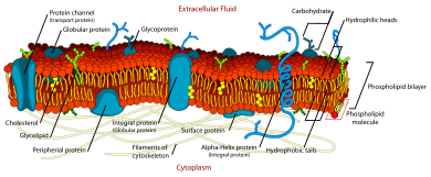
When taken together, the falsification evidence demonstrates that the Davson-Danielli Model, specifically around the proteins, was inaccurate. Singer and Nicolson presented the Fluid Mosaic Model in 1972. There have been a few adjustments with the discovery of cholesterol in animal cell membranes, but for the most part our current model of the cell membrane (see picture on right) remains very similar to what they purposed in 1972.
How is this model different from Davson-Danielli's Model?
|
https://upload.wikimedia.org/wikipedia/commons/thumb/d/da/Cell_membrane_detailed_diagram_en.svg/390px-Cell_membrane_detailed_diagram_en.svg.png

https://aworldofbiology.weebly.com/uploads/3/2/1/4/32143827/8830527_orig.jpg
|
|
|
|
Parts and Functions of the Cell Membrane

https://www.brainkart.com/media/extra2/hYw6bkV.jpg
https://study.com/cimages/multimages/16/fluidmosaic.png
|
|
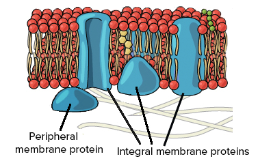
Membrane Bound Proteins
|
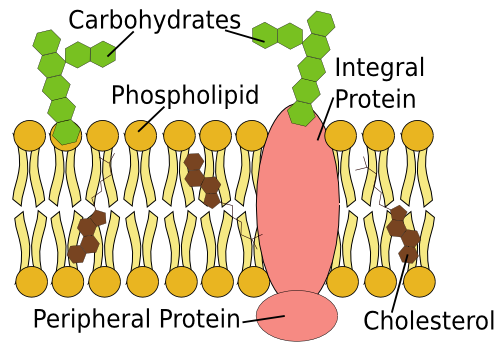
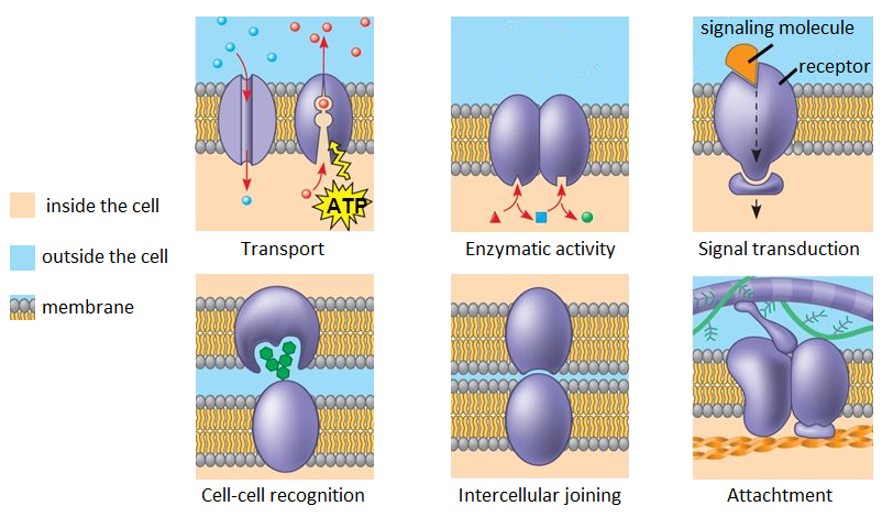
Membrane Proteins can be both integral (embedded across membrane) or peripheral (embedded on surface). They have a wide range of functions. Examples include:
|
https://cdn.kastatic.org/ka-perseus-images/234a6b91810a43e7e7ea3e9cbe8c60dee6ad3cee.png
https://d2jmvrsizmvf4x.cloudfront.net/g3p05qC0QeqJGCVTB5uW_plasmamebraneproteins.png
|
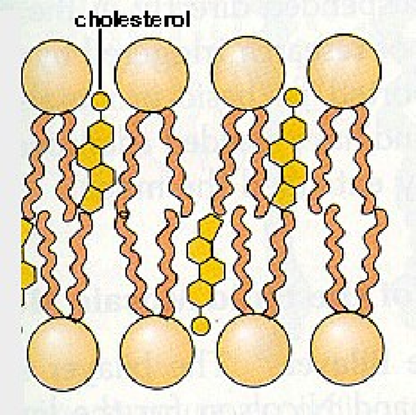
Cholesterol
https://www.researchgate.net/profile/Kumari_Ubhayasekera
/publication/27655102/figure/fig1/AS:310082899333135@1450940932640/
Position-of-cholesterol-among-phospholipid-molecules-in-cell-membrane.png
|

https://ib.bioninja.com.au/_Media/cholesterol-molecule_med.jpeg
Cholesterol: lipid (not a fat or oil), type of steroid.
|
